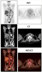
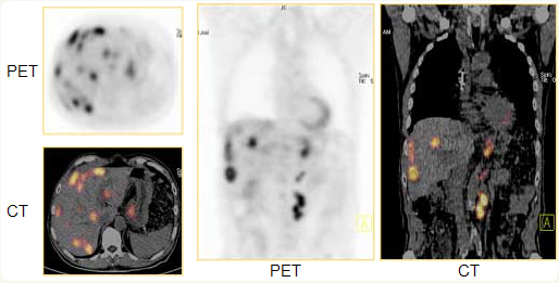
What Is a PET/CT Scan?
In one continuous full-body scan (usually about 30 minutes),  PET captures images of miniscule changes in the body’s metabolism caused by the growth of abnormal cells, while CT images simultaneously allow physicians to pinpoint the exact location, size and shape of the diseased tissue or tumor.
Essentially, small lesions or tumours are detected with PET and then precisely located with CT.
 Positron Emission Tomography (PET) and Computerized Tomography (CT) are both standard imaging tools that allow physicians to pinpoint the location of cancer within the body before making treatment recommendations.
The highly sensitive PET scan detects the metabolic signal of actively growing cancer cells in the body and the CT scan provides a detailed picture of the internal anatomy that reveals the location, size and shape of abnormal cancerous growths.
Alone, each imaging test has particular benefits and limitations but when the results of PET and CT scans are “fused” together, the combined image provides complete information on cancer location and metabolism. The bottom line is that you can have both scans – PET and CT – done at the same time.
How Does the Procedure Work?
While a CT scan provides anatomical detail (size and location of the tumor, mass, etc.), a PET scan provides metabolic detail (cellular activity of the tumor, mass, etc.). Combined PET/CT is more accurate than PET and CT alone! 1
 Anatomical: CT scanners send x-rays through the body, which are then measured by detectors in the CT scanner. A computer algorithm then processes those measurements to produce pictures of the body’s internal structures.
Metabolic: PET images begin with an injection of FDG, an analog of glucose that is tagged to the radionuclide F18. Metabolically active organs or tumors consume sugar at high rates, and as the tagged sugar starts to decay, it emits positrons. These positrons then collide with electrons, giving off gamma rays, and a computer converts the gamma rays into images. These images indicate metabolic “hot spots,” often indicating rapidly growing tumors (because cancerous cells generally consume more sugar/energy than other organs or tumors).
The entire examination usually takes less than 30 minutes, providing comprehensive diagnostic information to your health care team very quickly. The PET/CT system provides exceptional image quality and accuracy of diagnostic information.
What Does PET/CT See?
PET/CT scanning integrates PET and CT technologies into a single device, making it possible to obtain both anatomical and biological data during a single exam. This integrated approach permits accurate tumor detection and localization for a variety of cancers, including:
- Breast
- Esophageal
- Cervical
- Melanoma
- Lymphoma
- Lung
- Colorectal
- Head and Neck
- Ovarian
 PET/CT applications:
- Determines extent of disease
- Determines location of disease for biopsy, surgery or treatment planning
- Assesses response to and effectiveness of treatments
- Detects residual or recurrent disease
- May assist in avoiding invasive diagnostic procedure
Benefits of PET/CT
 There are tremendous benefits of having a combined PET/CT scan:
- Earlier diagnosis
- Accurate staging and localization
- Precise treatment and monitoring
With the high-tech images that the PET/CT scanner provides, patients are given a better chance at a good outcome and avoid unnecessary procedures. A PET/CT image also provides early detection of the recurrence of cancer, revealing tumors that might otherwise be obscured by scar tissue that results from surgery and radiation therapy, particularly in the head and neck.
In the past, difficulties arose from trying to interpret the results of a CT scan done at a different time and location than a PET scan, due to the fact that the patient’s body position had changed. The combination PET/CT provides physicians a more complete picture of what is occurring in the body – both anatomically and metabolically – at the same time
The Story of PET/CT
Doctors, especially cancer surgeons, were often frustrated in trying to match PET images with CT images to determine the precise location of a tumor in relation to an organ or the spinal column. They had little choice other than to “eyeball” the two separate images and make an educated guess as to the tumor’s exact location – until 1992, when engineer Ron Nutt and physicist David Townsend came up with the idea of combining a PET and CT into one machine.
After working on their combined PET and CT concept for three years, Nutt and Townsend received a grant from the National Cancer Institute. This enabled the completion of a prototype machine, which was installed at the University of Pittsburgh medical center in 1998.
The pair designed the machine to be more patient-friendly by making the diameter of the PET/CT tunnel 28 inches, far more spacious than the typical MRI tunnels.
Time Magazine honored PET/CT as the “Medical Science Invention of the Year” in 2000, noting that the PET/CT scanner has “provided medicine with a powerful new diagnostic tool.”
What Should I Expect?
Difficult questions deserve answers, and taking a “wait and see” approach is sometimes unacceptable. A PET scan helps answer those tough questions.
Your PET scan will produce a “picture” of how your body’s cells are functioning, whereas an x-ray, CT scan, or MRI produce a picture of bones, organs and tissues.
Your PET scan will help you and your physician make a more informed decision about your diagnosis and treatment path.
Some hints to prepare you for your scan:
- Dress comfortably and warmly. Scanner rooms can sometimes be cool.
- Avoid eating anything for at least four-six hours before your scan (this includes sugar-free gum, mints, candy and beverages other than water).
- No strenuous exercise the day of your exam (example: working out, jogging, etc.).
- Please bring a copy of your most recent CT, X-Ray, or MRI films with you on the day of your PET scan.
- Be prepared to lie still for 30-75 minutes while the scan is performed.
Procedure
A PET scan is completely painless and has no side effects. After fasting for 6 hours, you will receive an injection of a trace amount of radioactive glucose, which is distributed throughout the body.
About 30-75 minutes after the injection, you will empty your bladder, then lie down on a scanner bed. Images will be taken of your body as you lie still on the scanner bed.
How Long Will the Scan Take?
A scan takes approximately 1-2 hours, depending on the type of scan you are having (i.e., whole body, brain, etc.) and the type of scanner being used. The results are then interpreted by a trained nuclear medicine physician or radiologist and sent to your referring physician.
Diabetics
If you are diabetic, please call the facility for special instructions 080-22224050
After My PET Scan
When the PET scan is done, make sure to drink plenty of water or other fluids throughout the day.
Patient Instructions
Instruction for the patient planned to undergo PET CT scan
- 6-8 hours of fasting (longer the fasting better are the PET Scan results).
- Plenty of water is encouraged.
- In case of prolonged fasting or intolerance of being in empty stomach for longer time such patient’s can take cucumber or plane tomato juice only after such instructions from nuclear medicine task.
- Diabetic patients (usually are given appointment on priority basis in morning) need to personally ask the nuclear medicine desk for individualized approach.
- Pre menopausal patients need to inform the nuclear medicine doctor about their menstrual cycles if expected during the scheduled scan.
- Serum creatinine (required for CT scan) done with past 1 week of scheduled PET-CT scan.
- Patients scheduled for PET cardiac scan need to take a low carbohydrate and protein rich (egg, white, chicken and soy-bean) diet 2 days prior to the scan with 5 to 6 hours of fasting on the day of scan.
- Patients scheduled for PET brain scan need to be in quiet environment and strict 6 hours fasting in the morning of scheduled scan.
- Bring all your previous medical reports and CDs of previous PET scan (if done).
- Consult our radiation safety officer for the precautions to be taken during and after the procedure is over.
Our Services
-
PETCT Scan
In one continuous full-body scan (usually about 30 minutes), PET captures images of miniscule changes in the body’s metabolism caused by the growth of abnormal cells.
-
Nuclear Medicine
NM is a specialized form of medical imaging where small amount of radioactive isotopes are attached to molecules or chemicals which then form “Radiopharmaceuticals”.
-
SPECT
Myocardial perfusion imaging (MPI) is a non-invasive procedure used to test for significant coronary artery disease (CAD).
-
Radionuclide Therapy
Many radioisotopes, radio labelled conjugates are available for treatment of difficult medical conditions; Some of them are the treatment of choice for the given conditions.
-
Multi Slice CT
CT Scan or computed tomography is a painless and a non-invasive medical test that helps physicians diagnose and treat medical conditions.
Why choose us:
-
Latest in technology
-
High qualified consultants
-
Quick turn around
